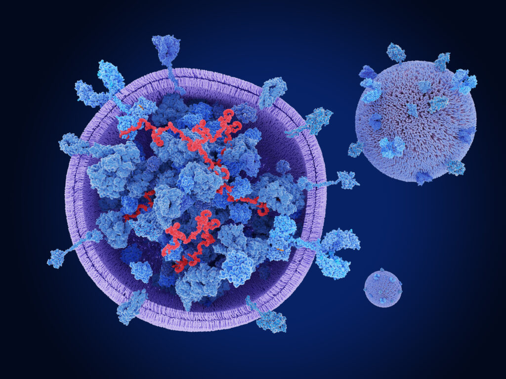
Doxorubicin (DOX) chemotherapy has long been recognized as an effective treatment for breast cancer, providing hope for many patients battling this formidable disease. However, as with many potent treatments, DOX comes with its own set of disadvantages, including significant side effects that can hinder its efficacy and patient quality of life. In recent years, the scientific community has turned its attention to exosomes (EXOs) as a potential means of enhancing drug delivery while minimizing adverse effects associated with conventional chemotherapy.
A groundbreaking study explored the use of EXOs as a vehicle for DOX, focusing on a co-culture system of malignant and non-malignant breast cells. This innovative in vitro model aimed to closely replicate the in vivo cellular microenvironment, allowing researchers to gain deeper insights into the treatment’s effects on both cancerous and healthy cells. The study was conducted by a team of researchers, including Moein Shirzad, Abdolreza Daraei, Hossein Najafzadehvarzi, Nazila Farnoush, and Hadi Parsian, all affiliated with the Babol University of Medical Sciences in Iran.
The researchers began by extracting extracellular matrices (EXOs) from mesenchymal stem cells derived from human adipose tissue through a process called ultracentrifugation. Following this, they employed techniques such as Western blotting, dynamic light scattering (DLS), and transmission electron microscopy to analyze the properties of the EXOs. Notably, DOX was encapsulated within the EXOs using sonication, and the loading rate was assessed using spectrophotometry.
In their experiments, the team assessed the cytotoxic effects of both free DOX and DOX encapsulated in EXOs (EXO-DOX) on various breast cell lines, including MCF-7, MCF-10A, MDA-MB-231, and A-MSC. The results were revealing: the methylthiazolyldiphenyl-tetrazolium bromide (MTT) assay determined that free DOX exhibited the highest cytotoxicity against MCF-10A cells, followed closely by MCF-7 cells. In contrast, EXO-DOX showed greater effectiveness against MCF-7 cells and demonstrated a lower half-maximal inhibitory concentration (IC50) compared to MDA-MB-231 cells.
The study also delved into the expression levels of various inflammatory cytokines, including IL-1β, IL-6, IL-10, and TNF-α. Free DOX was found to significantly downregulate the expression of pro-inflammatory cytokines—particularly in MCF-7 and MCF-10A cells—while simultaneously upregulating IL-10 expression. Remarkably, EXO-DOX induced a more pronounced alteration in cytokine expression compared to both the control and free DOX treatment groups.
One of the most compelling findings from this study was the observation of a synergistic effect of free DOX on cancer cells, which was paired with a reduction in the toxic effects of DOX on normal cells within the co-culture system. This dual action suggests that EXO-DOX could serve as a targeted drug delivery system, improving therapeutic efficacy while minimizing off-target toxicity.
The promising results of this research highlight the potential of using EXOs for targeted drug delivery in breast cancer treatment. As the scientific community continues to explore and refine this approach, the hope is that it may lead to safer and more effective therapies for patients battling breast cancer. The work of Shirzad, Daraei, Najafzadehvarzi, Farnoush, and Parsian represents a significant step forward in the quest to enhance cancer treatment while reducing the inherent risks associated with traditional chemotherapy.


