Cerebral ischemic stroke remains one of the most significant challenges in the realm of cerebrovascular diseases, standing as the leading cause of disability among adults. Recent advancements in medical research have opened doors to innovative therapeutic strategies. In this context, a study conducted by a team of researchers, including Yu-Sung Chiu, Kuo-Jen Wu, Seong-Jin Yu, Kun-Lieh Wu, Chang-Yi Hsieh, Yu-Sheng Chou, Kuan-Yu Chen, Yu-Syuan Wang, Eun-Kyung Bae, Tsai-Wei Hung, Shih-Hsun Lin, Chih-Hsueh Lin, Shu-Ching Hsu, Yun Wang, and Yun-Hsiang Chen, explores the potential benefits of human Wharton’s jelly mesenchymal stromal cells (WJ-MSCs) and their exosomes in combating the devastating effects of stroke.
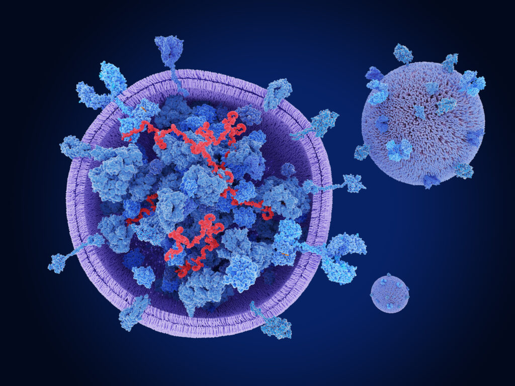
The study builds on prior research indicating that transplantation of WJ-MSCs can mitigate neuronal damage and foster functional recovery in animal models of stroke. This latest investigation delves deeper, specifically examining the effects of WJ-MSC exosomes (Exo) in both cellular and rat models of cerebral stroke. What sets this research apart is the focus on using exosomes—tiny vesicles that facilitate communication between cells—offering a non-invasive method for treatment.
The findings are promising. Administration of Exo significantly countered glutamate-induced neuronal loss in rat primary cortical neuronal cultures, demonstrating a protective effect against neuronal damage. In a controlled experiment, adult male rats underwent a 60-minute middle cerebral artery occlusion (MCAo) to mimic stroke conditions. After the procedure, Exo or a control vehicle was administered via tail vein injection shortly after the occlusion.
Behavioral assessments conducted two days post-treatment revealed remarkable improvements in the stroke-affected rats that received Exo. These rats exhibited enhanced locomotor function, greater forelimb strength, and reduced body asymmetry, as evidenced by lower Bederson’s neurological scores. These behavioral tests underscore the potential of Exo therapy in facilitating recovery post-stroke.
The researchers conducted further examinations of the brain tissues, which revealed a reduction in infarction volume among the animals treated with Exo. This was measured using 2,3,5-triphenyl tetrazolium chloride (TTC) staining, a standard method for assessing tissue viability in stroke research. Additionally, the transplantation of Exo was associated with an upregulation of neuroprotective factors such as BMP7 and GDNF, alongside anti-apoptotic factors like Bcl2 and Bcl-xL within the ischemic brain.
These results suggest that early post-treatment with WJ-MSC Exo not only contributes to improved functional recovery but also reduces brain damage following a stroke. The ease of non-invasive administration through the tail vein further enhances the appeal of this therapeutic approach.
As researchers continue to unravel the complexities of stroke recovery, the insights gained from this study represent a significant step forward. The potential for exosome-based therapies to mitigate the impacts of cerebral ischemic stroke may pave the way for new treatment protocols, offering hope to millions affected by this debilitating condition. The collaborative efforts of the authors, drawn from various institutions, underscore the importance of teamwork in advancing medical science and finding solutions to pressing health challenges.


